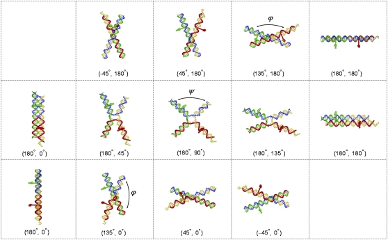
FIGURE 7.
Atomic-level representations of modeled HJ structures with the longest (20 bp) Cy3-Cy5 base separation, i.e., dyes on bases 10 bp from the recombination site. (Top) Coaxially stacked structures, generated by changes in ϕ, linking the antiparallel, lazy-H arrangement to the parallel form. (Middle) Kinked structures, obtained by variation of ψ, joining the two ideal antiparallel arrangements (shown in two views at the ends of the middle and upper/lower rows of images). (Bottom) Coaxially stacked structures, generated by changes in ϕ, linking the antiparallel upright-H configuration to the parallel form. DNA shown as a wireframe model with backbones represented by tubes (Cy3 donor-containing strand 3, green; Cy5 acceptor-containing strand 4, red; biotin-labeled strand 1, yellow; unlabeled strand 2, blue). Dyes depicted as space-filled forms with Cy3 in green and Cy5 in red. (ϕ, ψ) values denoted in parentheses below each image. Note the similar spacing of dyes and overall global fold of the partially kinked lazy-H structure and the nearby stacked-X arrangement where the (ϕ, ψ) values are (180°, 135°) and (135°, 180°), respectively.

