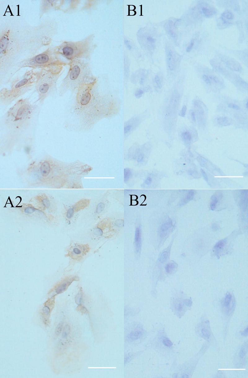
Figure 2.

Secondary cultures of newborn rabbit RPE cells expressed cytokeratin and S-100. RPE cells (passage 4) were plated on slides at a density of 5-10×104 cells per 1ml and cultured for 2 days. The cells were prepared for immunocytochemistry to determine whether the cells expressed RPE-specific cytokeratins using the monoclonal antibody MNF116 (A1) or S-100 using the monoclonal antibody of SH-B1 (B1). In panels (A2) and (B2), the primary antibody was omitted. The cells were counterstained with hematoxylin (blue) and observed by bright field. In each case, the brown immunostain was observed in virtually all of the cells. Bar, 25 μm.
