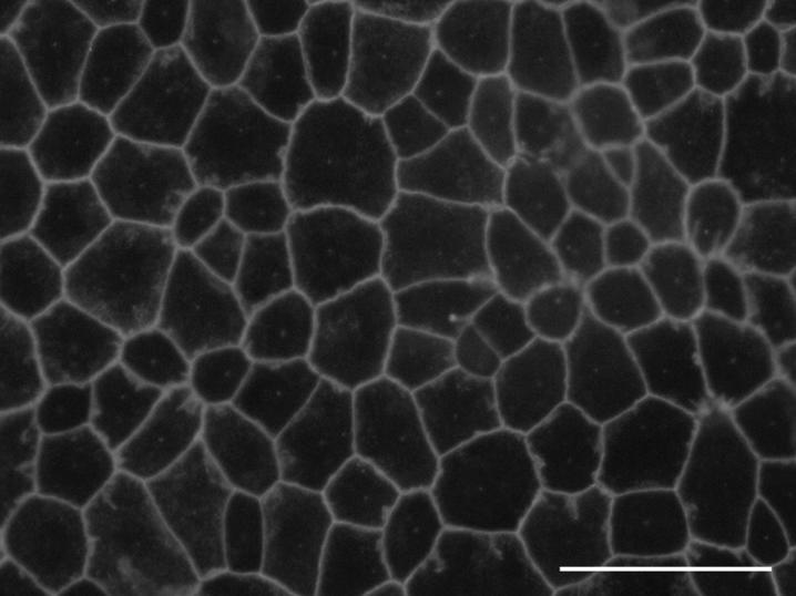
Figure 3.
ZO-1 distributed to circumferential bands where neighboring cells abutted in confluent cultures. Cells, freshly isolated from newborn rabbits, were cultured for 30 days on Transwell filters and stained for ZO-1 by indirect immunofluorescence. The ‘cobblestone’ appearance was typical of an epithelial monolayer. Bar, 50μm

