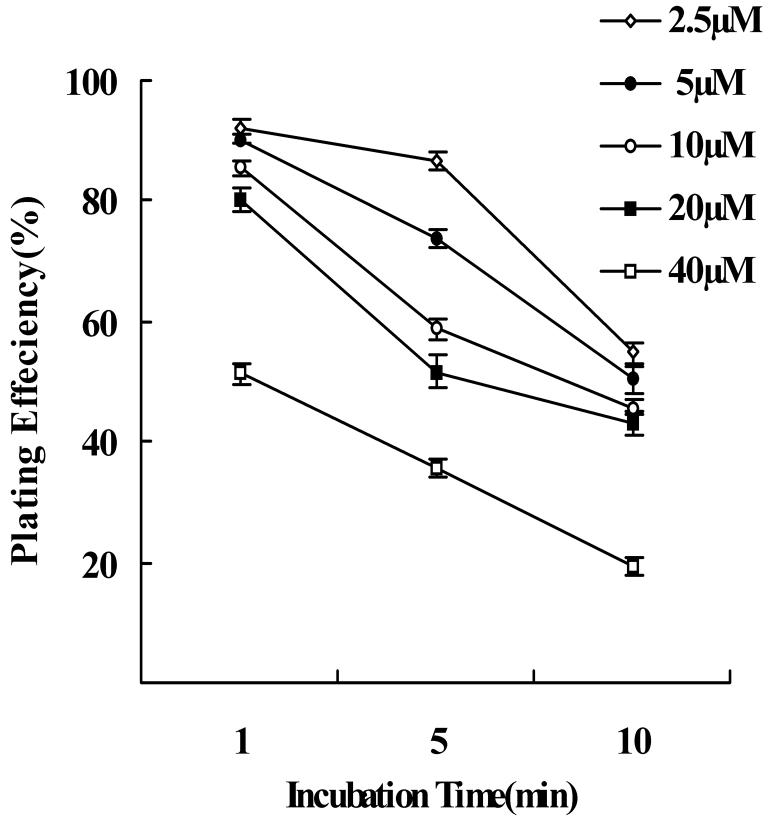
Figure 5.
Plating efficiency of newborn rabbit RPE cells following CFDA-SE labeling. The plating efficiency of RPE cells decreased with increasing incubation time in all concentrations of CFDA-SE. Following 10 minutes of incubation the plating efficiency decreased significantly at all dye concentrations. There was also significant decreased plating efficiency at concentrations 40μM compared to 2.5, 5, 10 and 20μM following 1 minute incubation (one-way ANOVA, P>0.05). Each point in the graph represents the mean percentage of plated RPE cells, after 24 hours. Error bars indicate the standard error.

