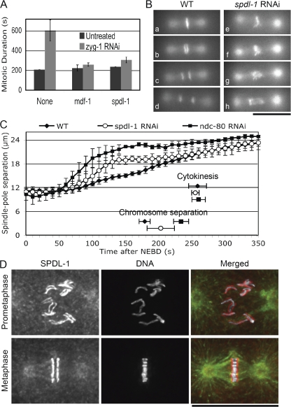
Figure 1.
SPDL-1 is required for proper chromosome segregation and SAC activation. (A) Mitotic duration from NEBD to chromosome decondensation was measured in AB cells of wild-type embryos dissected from adult hermaphrodites soaked with dsRNA of indicated genes alone (Untreated) or in combination with zyg-1 dsRNA (zyg-1 RNAi). Depletion of MDF-1 or SPDL-1 bypassed the ZYG-1 depletion–induced mitotic delay. (B) One-cell–stage embryos expressing GFP–histone H2B and GFP–tubulin were dissected before undergoing first mitosis from adult hermaphrodites injected with buffer (WT) or with spdl-1 dsRNA (spdl-1 RNAi) and studied by time-lapse fluorescence microscopy. Still images of embryos at 10 s before (a and e) and 0 (b and f), 20 (c and g), and 70 s (d and h) after the onset of anaphase (WT) or of anaphase-like separation of chromosome masses (spdl-1 RNAi) are shown. Bar, 20 μm. (C) Kinetics of centrosome separation in embryos dissected from wild-type adult hermaphrodites untreated (WT) or injected with dsRNA of indicated genes. Time at NEBD was set as 0 s. SPDL-1 depletion caused moderate acceleration of centrosome separation. Timing of chromosome separation and onset of cytokinesis are shown. Mean number of data obtained from at least three individual embryos are plotted and standard deviations are shown as error bars. (D) Immunofluorescence images of fixed wild-type embryos at indicated stages of the first mitosis. DNA (white), tubulin (green), and SPDL-1 (red) were stained with DAPI, anti-tubulin, and anti–SPDL-1 antibodies, respectively. SPDL-1 colocalizes to microtubules through mitosis and also temporarily localizes to kinetochores from prometaphase until anaphase. Bar, 20 μm.

