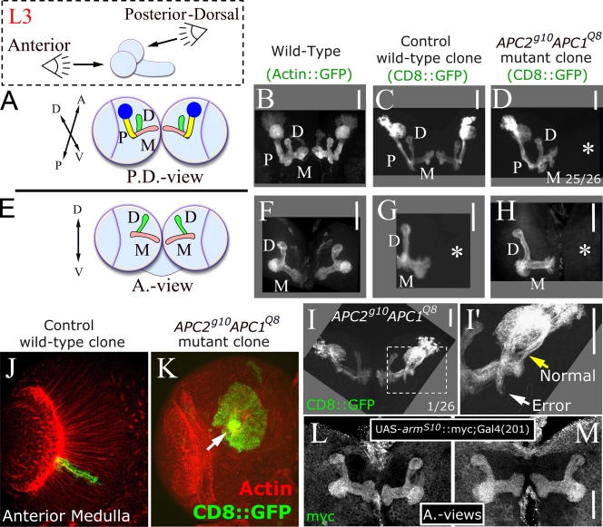
Figure 4.
APCs are not required for MB axon targeting. (A and E) Posterior-dorsal (A) and anterior views (E) of third instar MB. Cell body cluster plus dendritic region (blue), peduncle (P, yellow), and medial (M, pink) and dorsal (D, green) axon projections. (B and F) Wild type. (C and G) Control wild-type clones. (D and H) APC2g10APC1Q8 clones. Asterisks, brain hemispheres where a clone was not induced. (I and I′) Pathfinding error. (I′) Enlargement of area is shown in dashed square. White arrow indicates error, and yellow arrow indicates normal axon trajectory. (J and K) Wild-type and APC double null medullar neurons. Arrow indicates axon outgrowth defect. (L and M) MBs expressing armS10. Bars, 50 μm.

