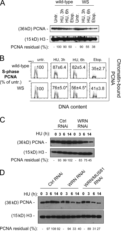
Figure 4.
Replication arrest induces dispersal of PCNA from S-phase chromatin in WRN-deficient cells. (A) Levels of chromatin-bound PCNA in wild-type and WS fibroblasts treated with 2 mM HU for the indicated times or with 50 μM etoposide for 6 h (Etop.). H3 histone was used as a loading control. The residual amount of PCNA in the chromatin fraction is expressed as the percentage of the amount in the untreated control normalized against the H3 histone content. (B) Levels of chromatin-bound PCNA in S-phase cells after replication inhibition. Wild-type and WS fibroblasts were exposed to 2 mM HU for the indicated times or to 50 μM etoposide for 6 h (Etop.), before being processed for biparametric PCNA/DNA flow cytometry. Representative cytograms for each condition are presented. The values shown represent the fluorescence intensity of the PCNA staining relative to an S-phase DNA content, expressed as percentage of the untreated control. The gates indicate the area considered for the evaluation of PCNA fluorescence intensity. *, statistically significant compared with the wild type; P < 0.01 (analysis of variance test). (C) Levels of chromatin-bound PCNA in HeLa cells transfected with control (GFP) or WRN siRNAs and treated with 2 mM HU for the indicated times. H3 histone was used as a loading control. The residual amount of PCNA in the chromatin fraction is expressed as the percentage of the amount in the untreated control normalized against the H3 histone content. (D) Levels of chromatin-bound PCNA in HeLa cells transfected with control (GFP), MUS81, and/or WRN siRNAs treated with 2 mM HU for the indicated times. H3 histone was determined as a loading control. RNAi-labeled samples represent cells treated with MUS81 siRNAs; if not otherwise specified, samples were treated with control siRNAs against GFP.

