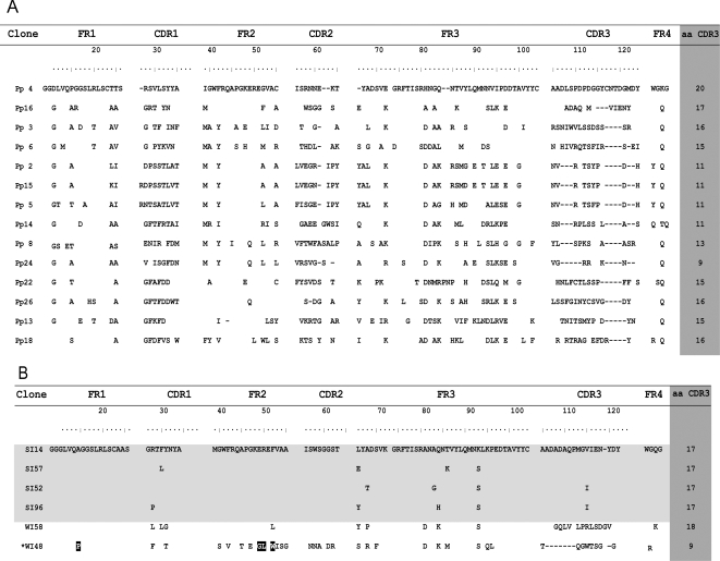
Figure 2.
(A) Alignment of the sequences of 14 randomly chosen clones from the library before panning (pre-panning clones are labeled pp) and (B) Sequence analysis of the six post-panning selected VHHs.The 4 strong inhibitors (SI) clones are shown in clear gray background. Lengths in CDR3 are shown in dark gray background. Clone WI48 is a conventional IgG (*) and its characteristic residues are marked in black. Spaces denote identical residues and dashes denote deletions. Numbering and CDR designations are according to IMGT numbering system [46].

