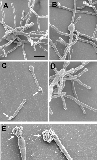
FIG. 7.
Scanning electron microscopy of hyphae grown at 48°C. Conidia of the different strains were seeded on glass coverslips and grown in AMM for 16 h at 48°C. (A) D141; (B) parental strain AfS35; (C) Δafmnt1 mutant; (D) complemented mutant Δafmnt1 afmnt1; and (E) higher magnification of the Δafmnt1 mutant. Arrows in panels C and E indicate amorphous material released at the hyphal tips. The bars in panels A and E represent 20 μm. The bar in panel A is also valid for panels B to D.

