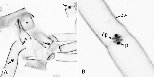
FIG. 2.
Confocal microscopy image (inverted) shows R. solani hyphal fragments stained with WGA conjugated to Alexa 488. Cell walls (cw), dolipore swellings (dp), and plugs (p) are labeled, whereas the septal plate hardly shows any fluorescence. Plugs are more intensely labeled than dolipore swellings and cell walls. (A) Overview; (B) detail image.

