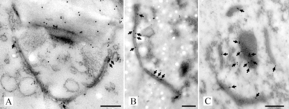
FIG. 6.
Transmission electron micrographs of high-pressure frozen, freeze-substituted, and Lowicryl HM20-embedded R. solani hyphae (A) and SPC-enriched fractions (B and C) labeled with anti-SPC18 antibodies. Antigen-antibody complexes were visualized with secondary goat-anti-rabbit antibodies conjugated with 10 nm gold. Gold particles (arrows) were located at the dolipore swelling and SPC in hyphal cells (A). Furthermore, sections of the SPC-enriched fractions showed gold label present at the SPC (B and C) and at the periphery of plug material that was connected to the SPC (C). Bars, 250 nm.

