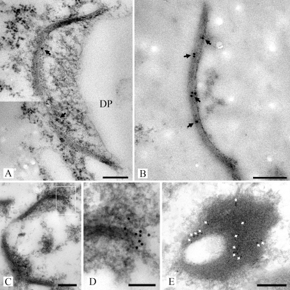
FIG. 8.
Transmission electron micrographs of high-pressure frozen, freeze-substituted, and Lowicryl HM20-embedded R. solani hyphae (A) and SPC-enriched fractions (B to E) labeled with anti-recSPC18 antibodies. Antigen-antibody complexes were visualized with secondary goat-anti-rabbit antibodies conjugated with 10 nm gold. Gold label (arrows) was present at the SPC (A to D). Gold label was preferentially localized at the base of the SPC (C and D). Panel D shows an enlargement of the box in panel C. Furthermore, label (white arrows) was preferentially localized at the periphery of the plug, as shown in this detailed image of a plug (E). Bars, 200 nm (A to C and E) and 100 nm (D).

