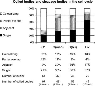
Figure 3.
Cells synchronized by mitotic shake-off (mso) and hydroxyurea (hu) treatment revealed that the spatial association between coiled bodies and cleavage bodies is cell cycle dependent. Coiled bodies were scored as colocalizing, partially overlapping, adjacent, or unassociated with a cleavage body (cleavage bodies were almost never seen unassociated with a coiled body). G1 cells were analyzed 4 h after mso; S phase cells were analyzed 10 h after mso or 4 h after release from hu block; and G2 cells were analyzed 8 h after release from hu block. These show that the nuclear bodies were mainly colocalizing in G1 and adjacent in S phase.

