Figure 6.
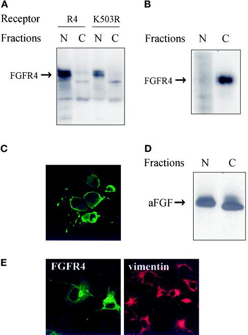
Characterization of the expressed receptor. (A) Western blot analysis of FGFRs in subcellular fractions of COS cells. Transfected COS cells were lysed and fractionated into a nuclear (N) and a cytoplasmic (C) fraction. The fractions were precipitated with trichloroacetic acid and analyzed by SDS-PAGE. Blots were probed with anti-FGFR4 antibodies. (B) Localization of surface-labeled FGFR4. COS cells transfected with FGFR4 were surface labeled by incubating them with Na125I, lactoperoxidase, and H2O2 for 5 min, and then DTT and tyrosine were added to stop the reaction. The cells were subsequently lysed and fractionated into a nuclear (N) and a cytoplasmic (C) fraction. The fractions were sonicated and centrifuged, and the receptors in the supernatants were immunoprecipitated with anti-FGFR4 antibodies. The immunoprecipitates were analyzed by SDS-PAGE and autoradiography. (C) Localization of detergent-insoluble FGFR4. Transfected COS cells were grown on coverslips and extracted in buffer containing 1% Triton X-100 before fixation with 3% paraformaldehyde. Fixed cells were incubated with antibody to FGFR4 followed by FITC-conjugated secondary antibody. (D) Nuclear and cytosolic fractions contain functional FGFRs. COS cells transfected with FGFR4 were lysed and fractionated into a nuclear (N) and a cytoplasmic (C) fraction. Both fractions were carefully sonicated for 5 s, and insoluble material was removed by centrifugation. Then the supernatants were incubated with [35S]methionine-labeled aFGF and 10 U/ml heparin for 3 h at 4°C, immunoprecipitated with anti-FGFR4 antibodies, and analyzed by SDS-PAGE and autoradiography. (E) Effect of transfection on the distribution of vimentin. Transiently transfected COS cells were fixed and double stained with anti-FGFR4 and anti-vimentin antibodies followed by FITC- and CY3-conjugated secondary antibodies, respectively.

