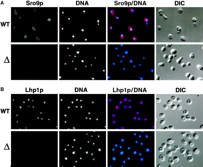
Figure 4.
Intracellular localization of Sro9p and Lhp1p. (A) Sro9p localizes to cytoplasmic dots. Anti-Sro9p antibodies were used to stain WT and sro9::URA3 cells and were visualized by indirect immunofluorescence microscopy. DNA was visualized by epifluorescence with Hoechst 33258. (B) Lhp1p localizes to the nucleus. WT cells and lhp1::LEU2 cells were stained with anti-Lhp1p antibodies and visualized as in A.

