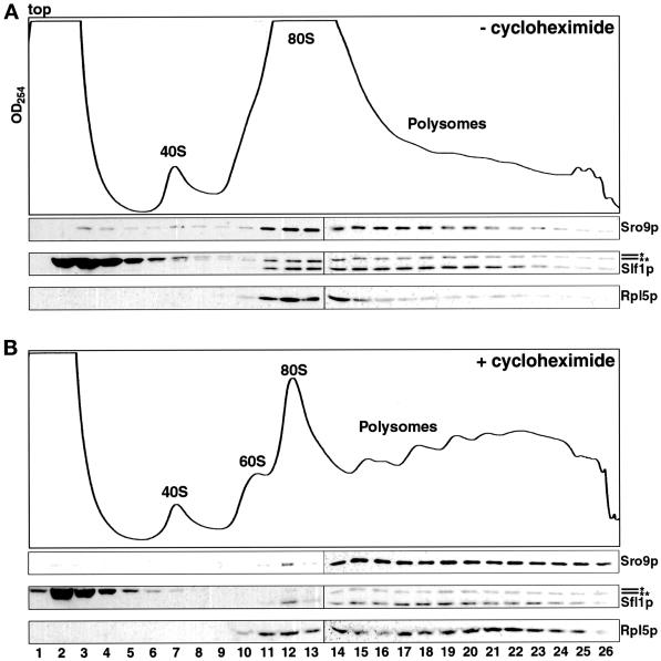
Figure 6.
Sro9p and Slf1p cosediment with translating ribosomes. Lysates were prepared in the absence (A) or presence (B) of cycloheximide. Supernatant (100 OD260 units of 10,000 × g) (see Figure 5) was applied to 20–47% linear sucrose gradients. After sedimentation, gradients were collected as the OD254 was monitored. The positions of 40S and 60S ribosomal subunits, 80S monosomes, and polyribosomes are indicated. Fractions were separated by SDS-PAGE, blotted, and probed sequentially with anti-Sro9p, anti-Slf1p, and anti-Rpl5p (which detects a ribosomal protein). Note the coincident shift in sedimentation of the ribosomes and Sro9p and Slf1p (A vs. B). The asterisk in the Slf1p immunoblots (*) denotes the unrelated background band (see Figure 3A). The double asterisk (**) denotes the Sro9p protein, because immunoblots were not stripped before reprobing. The vertical line in each immunoblot strip indicates that the strip was assembled from two separate immunoblots, which were processed and exposed in parallel.

