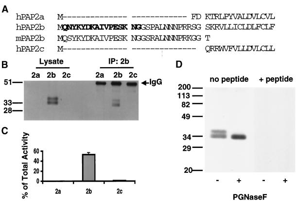
Figure 1.
Specificity and efficiency of the PAP2b antibody. (A) Sequence alignment of the N-terminal region of hPAP2a, hPAP2b, hPAP2c, and mouse PAP2b (expressed sequence tag AU03 6086). A peptide corresponding to residues 2–17 of the sequence of human PAP2b (letters in bold) was prepared and used to immunize rabbits as described in MATERIALS AND METHODS. (B) Fifty micrograms of total protein from TX100/βDOG lysates of baculovirus-infected Sf9 cells expressing hPAP2a, hPAP2b, or hPAP2c were subjected to immunoprecipitation using the anti-peptide PAP2b antibody. Ten micrograms of protein from lysates and one-twentieth of each immunoprecipitate (IP) were separated (12.5% SDS-PAGE), transferred, and analyzed for PAP2b by Western blotting. (C) PAP2 activity in the IP was determined and expressed as the percentage activity in the IP of the total PAP2 activity in the corresponding lysate (mean ± SD of triplicate determinations). (D) One hundred micrograms of protein from TX100/β-DOG lysates of baculovirus-infected Sf9 cells expressing hPAP2b were heated at 80°C for 3 min, then cooled on ice for an additional 5 min. Lysates were incubated in the presence or absence of PNGase F (1 U per assay) for 12 h at 37°C. Samples were boiled in sample buffer for 5 min to quench the reaction. Twenty micrograms of protein per lane were separated by SDS-PAGE (12.5%) and transferred to nitrocellulose membranes. Membranes were probed with PAP2b antibody or PAP2b antibody that was preabsorbed with 200-fold excess of the corresponding peptide.

