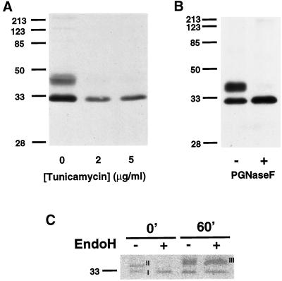
Figure 3.
Posttranslational modification of endogenous PAP2b in Swiss 3T3 Cells. (A) Swiss 3T3 cells (80% confluence) were grown in the presence of tunicamycin for 18 h at 37°C. Postnuclear supernatants (60 μg/lane) were prepared and analyzed for PAP2b by Western blotting. (B) Lysates were incubated with PNGase F (1 U per assay) for 12 h at 37°C as described in legend to Figure 1. Forty micrograms of protein per lane were separated by SDS-PAGE (12.5%) and analyzed for PAP2b by Western blotting. (C) Swiss 3T3 cells were labeled with 300 μCi per 100-mm dish of Trans-35S-Label (ICN) for 10 min, the media were removed, and cells were incubated in chase media containing 10 mM unlabeled methionine. PAP2b was immunoprecipitated, incubated with (+) or without (−) Endo H (10 mU per IP), and analyzed by SDS-PAGE (12.5%). A fluorogram of the gel is shown. Similar results were obtained in two additional experiments.

