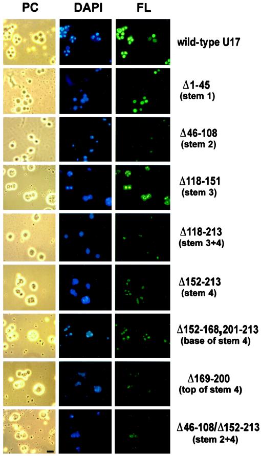
Figure 3.
Nucleolar localization of U17 snoRNA after deletion of sequences with stem structures and rRNA binding sites. Fluorescein-labeled U17 snoRNA was injected into the nuclei of X. laevis oocytes at an amount of 0.9 ng per oocyte. After 1.5 h, nucleolar preparations were analyzed by phase-contrast (PC) or fluorescence (FL) microscopy; the nucleolar rDNA was stained (DAPI, blue). U17 snoRNA carrying a deletion of stem 3 (Δ118–151) localized as well to nucleoli as the wild-type molecule (FL, green). U17 deleted in stem 1 (Δ1–45) localized strongly to nucleoli, and deletions of stem 2 (Δ46–108), stem 4 (Δ152–213), or a combination of both (Δ46–108/Δ152–213) showed significantly less but not abolished localization. Dissection of stem 4 (Δ169–200 or Δ152–168,201–213) did not reveal any major site of importance for nucleolar localization. The deletion of the entire structure of stems 3 and 4 between the single-stranded regions of conserved boxes H and ACA (Δ118–213) reduced but did not completely abolish nucleolar localization. Bar, 10 μm.

