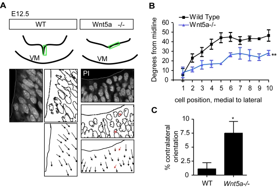
Figure 7. Apical-basal polarity/cell orientation are affected in Wnt5a−/− mice.
(A) At E12.5, propidium iodide staining on coronal sections through the VM revealed that cell nuclei in the neuroepithelium were rounded and their orientation was more variable with some cells pointing contralaterally (red asterisks/arrows). (B) The orientation of each cell nucleus was plotted versus its distance from the ventral midline. The angle between the nucleus and the midline was measured from cell 1 (the most medial) to cell 10 (the most lateral). Cell nuclei in Wnt5a−/− mice are oriented more ventrally compared to the more lateral orientation of cells in WT mice (two way-ANOVA for genotype and level, p = 0.0029, N = 3). (C) The frequency of cell nuclei oriented towards the contralateral ventral side (red arrows in A) was significantly increased in Wnt5a−/− mice (paired t-test, p = 0.0198, N = 3, 10 nuclei at 3 levels/animal).

