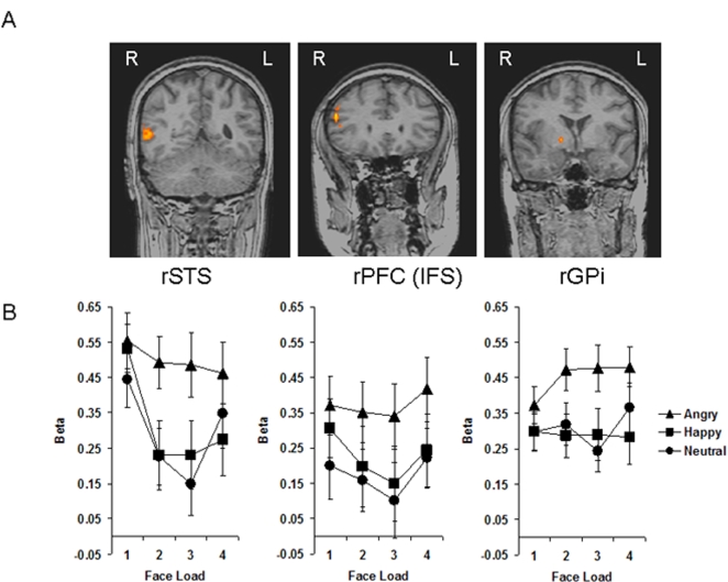
Figure 3.
(A) Three coronal brain slices show modulation of brain activity by emotional expression of faces in the VSTM task in the superior temporal sulcus (STS), prefrontal cortex (PFC) along the inferior frontal sulcus (IFS), and globus pallidus internus (GPi), all in the right hemisphere. (B) Beta values for each emotion and face load condition are plotted for the STS, PFC, and GPi. Activity is greater for angry vs. happy and neutral face expression conditions in all three brain regions. Bars represent±1 standard error.

