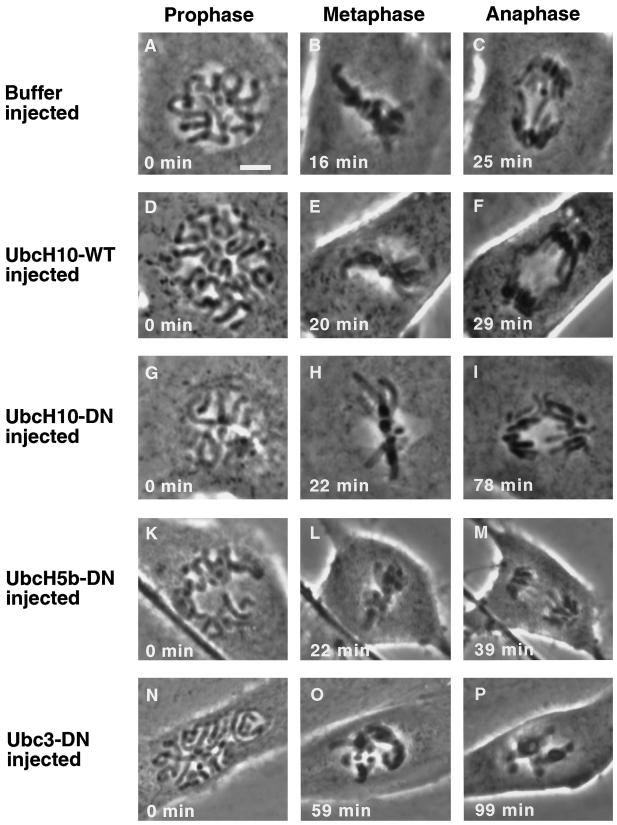
Figure 6.
Microinjection of purified dominant negative UbcH10 protein delays anaphase onset in PtK1 cells in vivo. Phase-contrast images are shown of PtK1 cells injected either buffer (A–C), wild-type UbcH10 protein (D–F), dominant negative UbcH10 (G–I), dominant negative UbcH5b (K–M), or dominant negative human Ubc3/CDC34 (N–P). All cells were injected in prophase (2–6 min before capture of the image), and time was determined when chromosomes aligned at the metaphase plate (metaphase) and separated subsequently (anaphase). Bar, 5 μm.

