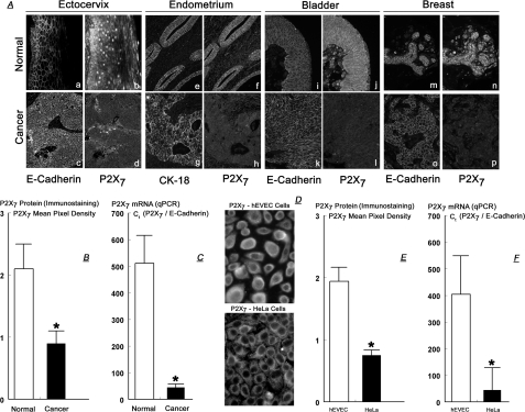
FIGURE 2.
A, tissue cross-sections from four patients with ectocervical squamous carcinoma (panels a–d), endometrial adenocarcinoma (panels e–h), bladder transitional cell carcinoma (panels i–l), and breast ductal adenocarcinoma (panels m–p). Tissues were co-immunostained with anti-P2X7 plus anti-E-cadherin or with anti-P2X7 plus anti-CK-18 antibodies (10 times). Data are representative of a total of 12 similar cases. B, semiquantitative analysis of P2X7 immunostaining in four cases of ectocervical cancers. C, qPCR data of P2X7 mRNA (normalized to E-cadherin mRNA) in normal (n = 4) and cancer (n = 3) ectocervical tissues. D, immunostaining with anti-P2X7 antibody of cultured hEVEC and HeLa cells (20 times). E, semi-quantitative analysis of P2X7 immunostaining in hEVEC and HeLa cells. F, qPCR data of P2X7 mRNA (normalized to E-cadherin mRNA) in hEVEC and HeLa cells. Experiments D–F were repeated five times. B, C, E, and F, *, = p < 0.001 compared with normal tissues or hEVEC cells.

