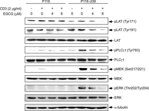
FIGURE 4.
The effect of EGCG on CD3-induced phosphorylation of downstream kinases. Cells (P116 and P116.cl39) were starved for 14 h and then incubated for 1 h with EGCG at various concentrations (0, 4, or 8 μm). Cells were washed and then stimulated with 2 μg/ml of CD3 for 30 min. Thirty μg of cellular extract were separated in each lane on a 10% SDS-polyacrylamide gel as described under “Experimental Procedures.” Equal protein loading and transfer of proteins were confirmed by stripping and incubating the same membrane with anti-α-tubulin.

