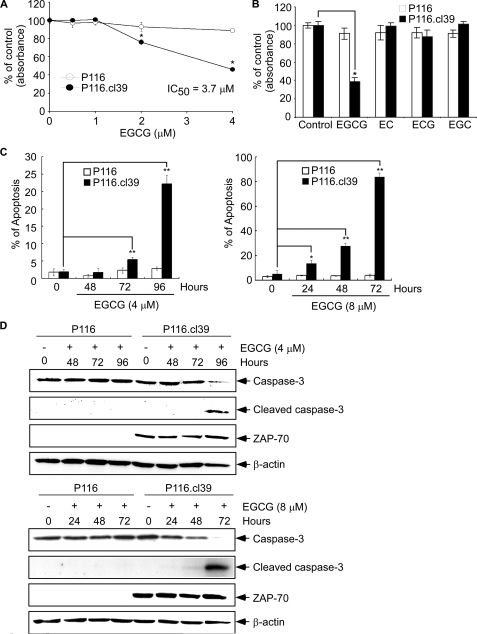
FIGURE 6.
The effect of EGCG and EGCG analogs on cell viability and apoptosis in P116 and P116. cl39 cells. A, effect of EGCG on cell viability. P116 or P116.cl39 cells (1 × 10³ cells/200 μl) were each seeded in a 96-well microtiter plate and incubated for 72 h with increasing concentrations of EGCG. The effect of EGCG on cell viability was estimated with the MTS assay kit, as described under “Experimental Procedures.” B, specific effect of EGCG on cell viability was compared with EGCG analogues in P116 and P116.cl39 cells by the MTS assay. C, analysis of apoptosis by flow cytometry after 4 μm EGCG (left) or 8 μm EGCG (right) treatment of cells for various times. Cells were labeled with annexin V-FITC and propidium iodide, and the distribution pattern of live and apoptotic cells was determined by flow cytometry. The graphs indicate percentage of early apoptotic and late apoptotic/necrotic cells. EGCG-treated cells were compared with untreated cells, and data are shown as the average of triplicate samples from three independent experiments. The asterisks (*, p < 0.005; **, p < 0.0001) indicate a significant increase in apoptosis in EGCG-treated cells compared with untreated cells. D, effect of EGCG on expression of caspase-3 and cleaved caspase-3 in P116 and P116.cl39 cells as determined by Western blotting.

