FIGURE 4.
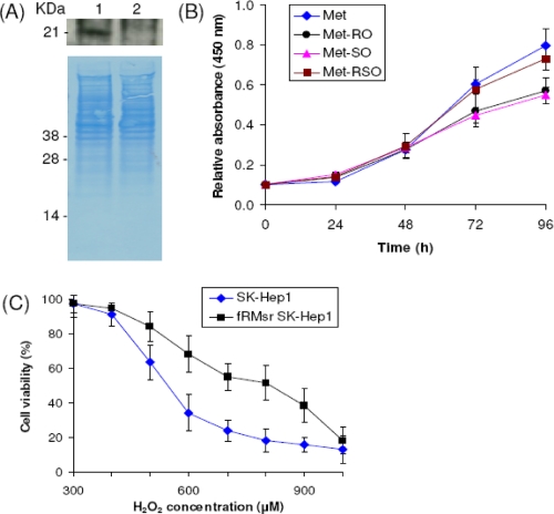
Characterization of SK-Hep1 cells expressing yeast fRMsr. A, Western blot analysis of SK-Hep1 cells stably expressing yeast His-tagged fRMsr (lane 1) and control SK-Hep1 cells (lane 2) with anti-His antibodies. B, yeast fRMsr-transfected SK-Hep1 cells were grown in media containing 0.1 mm Met (diamonds), 0.1 mm Met-RO (circles), 0.1 mm Met-SO (triangles), or 0.1 mm Met-RSO (squares) until 96 h. Cell growth was measured by an MTS cell proliferation assay at 0, 24, 48, 72, and 96 h. C, resistance of fRMsr-expressing SK-Hep1 cells to hydrogen peroxide treatment. Control (diamonds) and fRMsr-expressing SH-Hep1 (squares) cells were treated with the indicated concentrations of hydrogen peroxide, and their viability was assayed at 24 h. The error bars show the standard deviations from three independent experiments.

