FIGURE 4.
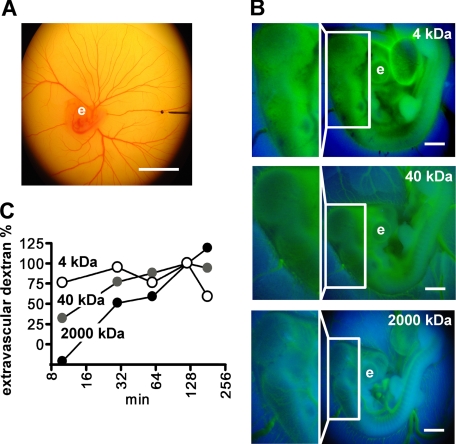
Microperfusion of chicken embryos and assessment of vascular permeability. A, a glass pipette filled with Trypan Blue is positioned in a vitelline vein (80 μm in diameter) for microperfusion of a 3-day-old embryo. Scale bar, 10 mm. B, dextran-FITC of different molecular mass was microper-fused into embryos and fluorescence images (excitation wave length, 485 nm) were taken at different times for quantitation of extravasated dextran. A representative set of images of the embryos 10 min after injection of dextrans is shown. The boxed areas are also shown at higher magnification. Note that 4-kDa dextran extravasates at the earliest time point observed. e, eye; scale bar, 1 mm. C, assessment of vascular permeability. The time course of extravasation of the different dextrans was assessed by image analysis and is shown relative to the value reached after 120 min. Representative data from three independent experiments are shown.

