FIGURE 3.
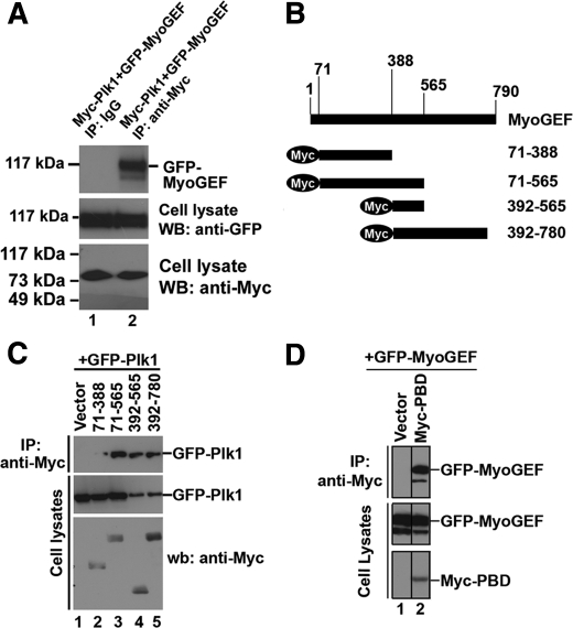
Interaction between MyoGEF and Plk1. A, HeLa cells were transfected with plasmids encoding Myc-Plk1 and GFP-MyoGEF. Anti-Myc-conjugated agarose was used to precipitate Myc-Plk1 from the transfected cell lysate, followed by immunoblotting with an antibody specific for GFP. B, schematic diagram of MyoGEF fragments. The numbers indicate the amino acids. C, a plasmid encoding GFP-Plk1 was cotransfected into HeLa cells with empty vector or plasmids encoding Myc-tagged MyoGEF fragments (71–388, 71–565, 392–565, or 392–780). Anti-Myc-conjugated agarose was used to precipitate Myc-tagged MyoGEF fragments from the transfected cell lysate, followed by immunoblotting with an antibody specific for GFP. D, a plasmid encoding GFP-MyoGEF was cotransfected into HeLa cells with empty vector or a plasmid encoding Myc-PBD. Anti-Myc-conjugated agarose was used to precipitate Myc-PBD from the transfected cell lysate, followed by immunoblotting with an antibody specific for GFP. wb, Western blot; IP, immunoprecipitation.

