FIGURE 6.
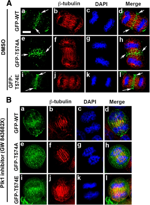
Localization of GFP-tagged MyoGEF at the central spindle is sensitive to Plk1 activity. A, localization of GFP-tagged MyoGEF at the central spindle. HeLa cell were transfected with plasmids encoding GFP-MyoGEF-wt (GFP-WT; panels a–d), GFP-MyoGEF-T574A (GFP-T574A; panels e–h), or GFP-MyoGEF-T574E (GFP-T574E; panels i–l). Arrows indicate the central spindle. B, the transfected cells in A were cultured in the presence of Plk1 inhibitor GW 843682X at 1 μm for 30–40 min (41). The treated cells were fixed in 4% paraformaldehyde and processed for immunofluorescence with an antibody specific for β-tubulin (red). The nuclei were stained with DAPI (blue). Bar, 20 μm. DMSO, dimethyl sulfoxide.

