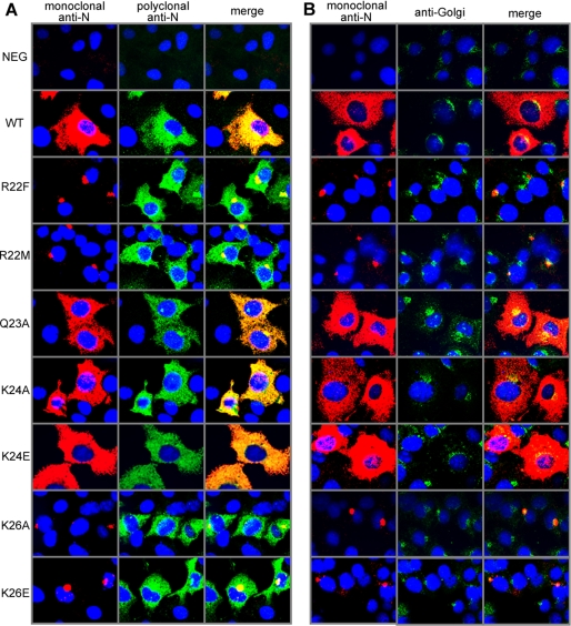
FIGURE 4.
Immunocytochemistry of full-length N protein with point mutations in the N1–74 coiled coil domain. Cos-7 cells were transfected with a plasmid expressing Andes virus N protein. Two days after transfection, the cells were fixed for immunofluorescence microscopy and double labeled with monoclonal (red) and polyclonal (green) anti-nucleocapsid antibodies (A) and monoclonal anti-nucleocapsid antibody (red) and anti-Golgi antibody (green) (B). The cell nuclei were stained blue using 4′,6-diamidino-2-phenylindole. Point mutations in Arg22 and Lys26 showed a dramatic difference in the monoclonal antibody recognition of Golgi-associated N protein, suggesting that the conformation or molecular interaction (or both) of the N protein is different when it is in the cytoplasm or when it is associated with the Golgi. WT, wild type.

