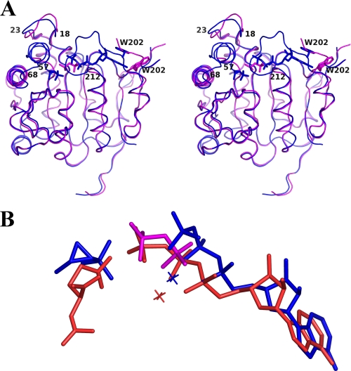
FIGURE 4.
A, a stereo view of the superposition of FomA·DPO (magenta) and the FomA·MgAMPPNP·fosfomycin (blue) structures. The polypeptide chains are shown as ribbon representations, AMPPNP, DPO, and fosfomycin molecules are shown in stick representation. B, a superposition of the bound substrates in the FomA·DPO (magenta), FomA·MgAMPPNP·fosfomycin (blue), and NAGK·MgAMPPNP·NAG (orange) complexes. Mg2+ cations are shown as crosses.

