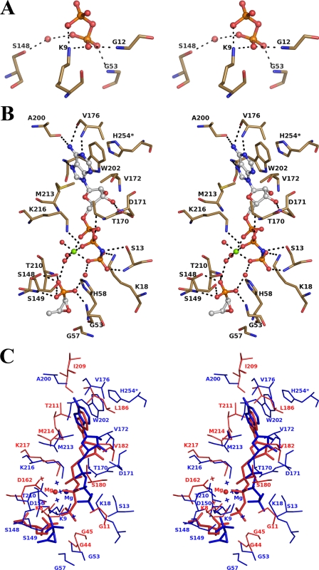
FIGURE 5.
Stereo views of the active site. A, FomA·DPO complex. B, FomA·MgAMPPNP·fosfomycin complex. The ligand molecules are shown in ball-and-stick format. The Mg2+ (green) and coordinated water molecules are represented as spheres. The interacting protein residues are shown in stick format. C, a stereo view of the superposition of the AMPPNP binding sites in FomA·MgAMPPNP·fosfomycin (blue) and NAGK·MgAMPPNP·NAD (red) structures. Mg2+ cations are shown as spheres, and water molecules are shown as crosses.

