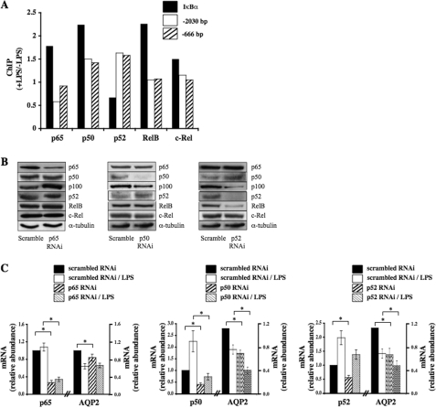
FIGURE 5.
The effect of LPS is dependent on altered p65, p52, and p50 binding to the AQP2 promoter. A, cells were challenged or not with 100 ng/ml LPS for 3 h before DNA fragmentation and immunoprecipitation using anti-p65, p50, p52, RelB, or c-Rel antibodies. Real-time PCR was performed using primers flanking either the NF-κB binding element of the AQP2 promoter (–2030, open box; –666, hatched box) or using primers flanking NF-κB binding element of the IκBα promoter (closed box). Results are expressed as the ratio of values obtained in the presence and in the absence of LPS. Shown are results of one of three similar experiments. ChIP, chromatin immunoprecipitation. B and C, cells were transfected with either scrambled RNAi or RNAi targeting various NF-κB isoforms and then challenged or not with 100 ng/ml LPS for 3 h before protein (B) or RNA (C) extraction. B, Western blot analysis was performed on various NF-κB isoforms and on α-tubulin, used as a loading control. Representative images are shown. C, real-time PCR from RNA extracts of cells transfected with scrambled RNAi or RNAi against p65 (left panel), p50 (middle panel), or p52 (right panel) was performed using primers specific for NF-κB isoforms or AQP2. Results are expressed relative to control values determined in cells transfected with scrambled RNAi and subjected to isotonic medium. Bars are the mean ± S.E. from six independent experiments. *, p < 0.05.

