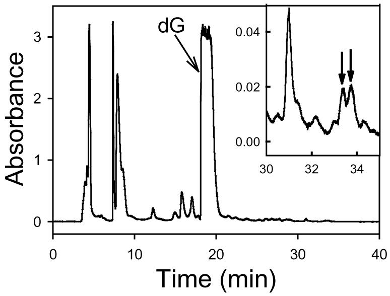
Figure 1.

The HPLC chromatogram of the reaction mixture of P5D and dG incubated at pH 7.4 and 37 °C for 426 h (monitoring wavelength: 260 nm). Inset: The magnified chromatogram between 30 and 35 min to show the minor peaks. The two peaks, which were not observed in the chromatograms of P5D and dG controls, are marked with arrows.
