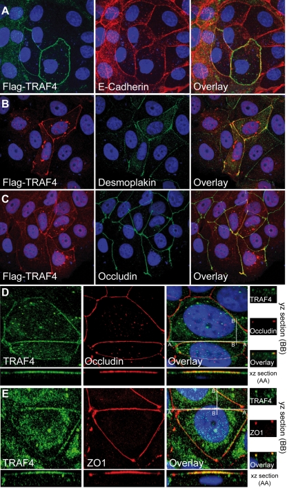
Figure 4. TRAF4 localization at cell-cell junctions.
(A–C) : MCF7 cells transiently transfected with Flag-TRAF4 were subjected to IF experiments. Co-staining was performed using an anti-Flag antibody in combination with either anti-E-cadherin (panel A), anti-desmoplakin (panel B), or anti-occludin (panel C) antibodies specific for AJs, desmosomes, and TJs, respectively. A partial colocalization of TRAF4 with the 3 proteins was observed, the highest overlapping rate was observed with occludin. (D) : Co-staining of endogenous TRAF4 and occludin in MCF7 cells using the 2H1 anti-TRAF4 and anti-occludin antibodies. xz- and yz-scans show the expected linear and punctual localization of human TJ-related protein occludin (red) in sections of 0.4 µm from the apical pole of the cell. TRAF4 (green) exhibited similar staining. (E) : Co-staining of endogenous TRAF4 (green) and TJ-related protein ZO1 (red) in MCF7 cells using the 2H1 anti-TRAF4 and anti-ZO1 antibodies. There is a perfect colocalization of these proteins. Collectively, these data show that, at the plasma membrane, TRAF4 is restricted to the TJs. For all experiments, nuclei were stained using Hoechst-33258 (blue), and microscopy analysis was performed using a Zeiss Axiovert 100 M confocal laser scanning microscope equipped with LSM5 Pascal (Jena, Germany). The figure represents image projection of several confocal sections. Magnification : A–C , 40×; D and E, 63×.

