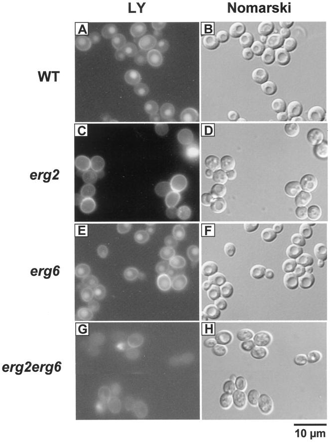
Figure 3.
Analysis of fluid phase endocytosis in ergΔ mutant cells. Wild-type (RH448; A and B), erg2Δ (RH2897; C and D), erg6Δ (RH3622; E and F) and erg2Δerg6Δ (RH3616; G and H) cells were incubated with LY at 24°C. To visualize the localization of LY, cells were viewed by FITC-fluorescence optics (A, C, E, and G). The same fields of cells were examined by Nomarski optics to visualize the vacuoles (which are apparent as indentations in the cell profiles; B, D, F, and H). Bar, 10 μm.

