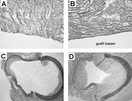
Figure 2.
Representative sections obtained at four weeks after arteri-ovenous graft implantation in pigs. Enhanced endothelialization in the anti-CD34-coated grafts coincided with profound increase in intimal hyperplasia at the venous anastomosis. Lectin-stained sections of bare (A) and CD34-coated grafts (B) obtained from the centre of the graft. Lectin is a marker for endothelial cells. Elastin von Gieson-stained sections of the venous anastomosis of bare grafts (C) and CD34-coated grafts (D). Reproduced from reference 11 with permission

