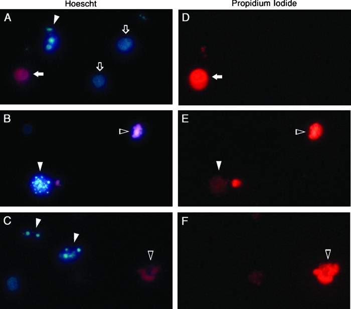
Figure 2.

HL-60 cells displaying representative patterns of fluorescent staining with Hoescht and propidium iodide following induction of apoptosis with VP-16. Cells were stained with both Hoescht 33342 and propidium iodide and visualized for Hoescht fluorescence (A–C) and propidium iodide fluorescence (D–F) with pairs of panels (A and D, B and E, and C and F) representing three fields of view (original magnification, 400×). Chromatin of healthy cells stains homogeneously with Hoescht (open arrows; A) but not at all with propidium iodide (D). Cells with condensed chromatin but intact plasma membranes stain strongly with Hoescht (filled arrowheads; A and C) but not at all with propidium iodide (D and F), illustrating the very early stages of apoptosis. A cell further along the apoptotic program displays condensed chromatin with Hoescht (filled arrowhead; B) and slight uptake of propidium iodide (filled arrowhead; E), suggesting decreased membrane selectivity. As apoptosis progresses to completion, cells with condensed chromatin (open arrowheads; B and C) also stain intensely with propidium iodide (open arrowheads; E and F), indicating profound disruption of plasma membrane integrity. Less frequent necrotic cells are easily distinguished from healthy or apoptotic cells by homogeneous chromatin staining with both Hoescht (filled arrow; A) and propidium iodide (filled arrow; D).
