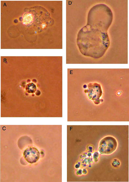
Figure 3.

Phase-contrast microscopy of HL-60 cells showing membrane blebbing after exposure to etoposide. Six hours after students exposed HL-60 cells to VP-16, cells with membrane blebbing were visualized using a phase-contrast microscope and photographed with a digital camera. Original magnifications: B, C, E, and F, 400×; A and D, 1000× (oil immersion).
