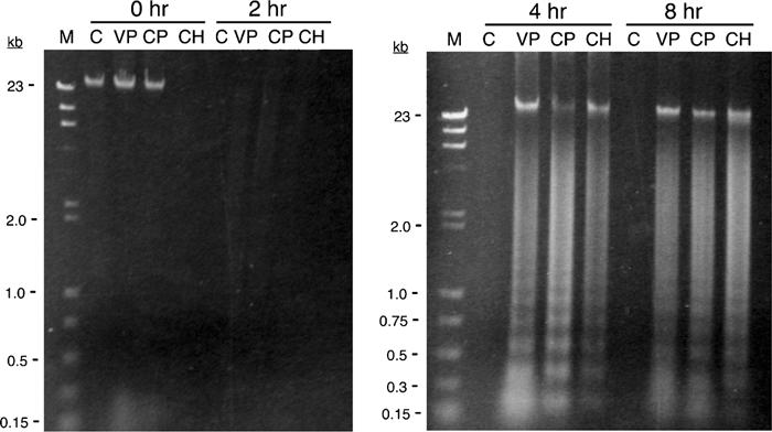
Figure 7.

Student-generated DNA laddering in HL-60 cells exposed to three apoptosis-inducing agents. HL-60 cells were exposed to either DMSO alone (C), 500 μM VP-16 (VP), 11 μM camptothecin (CP), or 4 μM cycloheximide (CH) for the indicated times. Cells were lysed, and the DNA was isolated by phenol–chloroform extraction followed by ethanol precipitation. The DNA was resolved by electrophoresis through a 1.2% TAE–agarose gel and visualized with ethidium bromide. The relevant sizes of the standard markers (M) are shown as kilobases (kb). DNA laddering is clearly evident within 4 h after exposure to all three inducers. Note, however, that several DNA samples, especially at the 2-h time point, were likely lost during the isolation procedure.
