
Fig. 3.
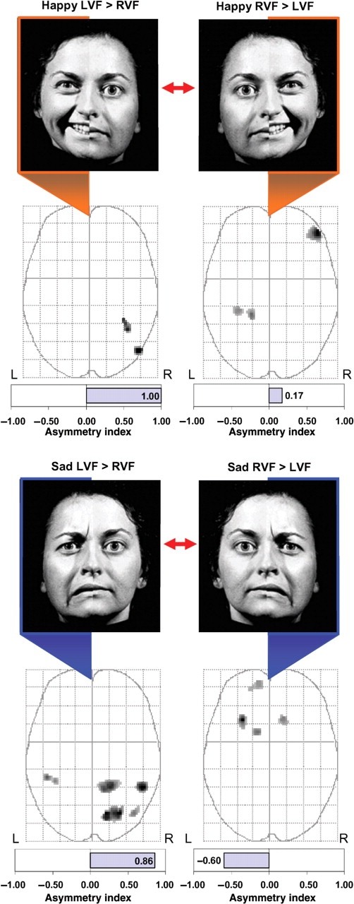
Comparisons between lateralized stimulus conditions. Brain activity associated with unilateral LVF presentations was compared directly to activity associated with unilateral RVF presentations using paired t-tests. Regions showing significant differences (P < 0.005, uncorrected) between the two lateralized presentation conditions are displayed on the MIPs in the axial plane. Happy hemifaces presented to the LVF produced significantly greater activity in the posterior right hemisphere (fusiform gyrus and middle temporal gyrus) than when presented to the RVF (top left). In contrast, happy hemifaces presented to the RVF produced greater activity in the right lateral orbitofrontal cortex and left fusiform/parahippocampal gyrus (top right). For sad hemifaces, LVF presentations produced greater activity predominantly in the posterior right hemisphere compared to similar RVF presentations (bottom left). In contrast, RVF presentations produced greater activity in the left orbitofrontal cortex and basal ganglia when compared to identical LVF presentations (bottom right).