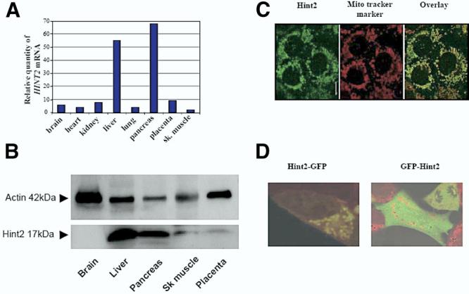
Figure 2.

(A) Tissue distribution of HINT2 mRNA assessed by real-time quantitative PCR using a human cDNA panel (Clontech). Results were normalized to GAPDH expression and presented as fold variations comparatively with skeletal muscle. (B) Expression of Hint2 protein in different human tissues assessed by immunoblot analysis with anantibody against Hint2. (C) Immunocytochemistry for Hint2 in Huh-7 cells. Hint2 (in green, left panel, bar = 10μm) colocalized with the mitochondrial marker Mito Tracker (in red, middle panel) as shown in the overlay (in yellow, right panel). (D) Overlay confocal microscopy pictures of HEK-293 cells incubated with Mito Tracker to mark the mitochondria in red and expressing a chimeric protein made of Hint2 and GFP. When the GFP was at the C-terminus of Hint2, Hint2 localized to the mitochondria (left panel). When the GFP protein was at the N-terminus of Hint2, Hint2 remained in the cytoplasm (right panel; original magnification 40×).
