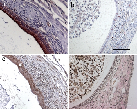
Figure 3:
Histological and immunohistochemical examination of EGFP-expressing endometriotic lesions.
(a) Anti-CK staining shows glandular epithelial cells. (b) Proliferating cells positive for Ki67 in cyst epithelium, stroma and in cyst content display red stained nuclei. (c) Estrogen receptor alpha positive cells in epithelium and stroma are indicated by brown stained nuclei. (d) W-E-vG staining of an endometriotic lesion reveals collagen connective tissue in red and nuclei in black. Scale bar: a, c, d = 50 µm, b = 100 µm. Original magnification: a, c, d = ×200, b = ×100.

