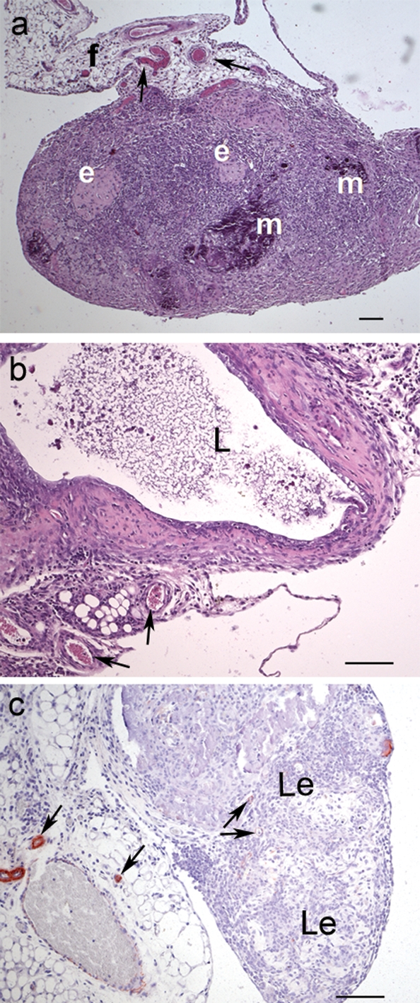
Figure 5:

Histological examination of lesions.
(a) Haematoxylin–eosin stained lesion from a TM-treated mouse with compact content, showing supporting vessels (indicated by arrows) embedded in fat (f), several sites of mineralization (m) and eosinophile patches (e). (b) Lesion from a control mouse with cystic lumen (L) surrounded by stromal cells and supporting vessels. (c) Immunohistochemical smooth muscle actin staining reveals small vessels in the supporting fat tissue and a few pericytes in the lesions (Le), indicated by the arrows. No further actin staining can be detected. Scale bars: 100 µm. Original magnification: a = ×50, b–c = ×100.
