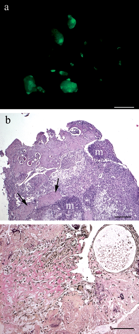
Figure 6:

Non-attached debris of a TM-treated mouse removed after autopsy.
a) Debris under fluorescent light in a TM-treated mouse. (b) Haematoxylin–eosin staining of a paraffin-embedded debris section. Eosinophil patches are indicated by arrows. Mineralized tissue (m) demonstrates ongoing necrosis. (c) W-E-vG staining of a debris section. Collagen connective tissue stains red, muscle yellow and nuclei black. Scale bar: a = 2 mm, b = 200 µm, c = 100 µm. Original magnification: b = ×50, c = ×100.
