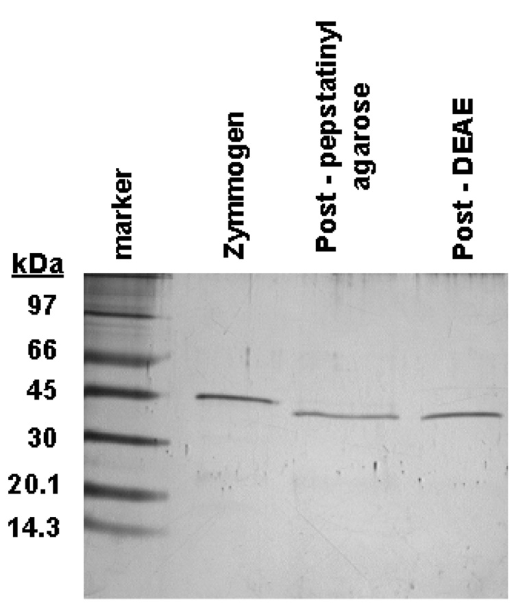
Figure 3.
SDS-PAGE analysis of purification of refolded "short" rHuCatD from the refolding mixture. The far left lane corresponds to molecular weight markers. Lane #1 represents full length rHuCatD from the refolding mixture Lane #2 shows the pepstatinyl-agarose fraction. Lane #3 illustrates the ion-exchange purified “short” pseudo rHuCatD migrating to about 42 kDa. The single band indicates purity to near homogeneity as judged by silver staining.

