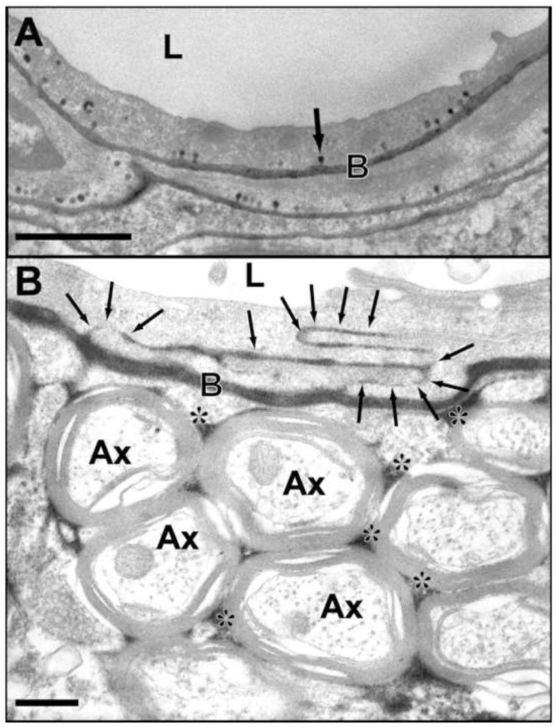
FIGURE 8.

Electron micrographs showing transcellular and intracellular passage of horseradish peroxidase (HRP) after ultrasound-induced BBB disruption. This tracer appears black in the photomicrographs and has a molecular weight of 40 kDa. A: Intense vesicular transport is observed by numerous HRP-positive caveolae (example indicated by arrow) after sonication at 0.26 MHz in a rabbit brain. B: Passage of HRP through several interendothelial clefts (arrows) is observed after sonication at 1.5 MHz in a rat. The tracer has infiltrated the basement membrane and the interstitial space in the neuropil (*). (L: lumen; Ax: cross-sectioned axons; B: basement membrane). (Data from [201, 202]; Courtesy of N. Sheikov).
