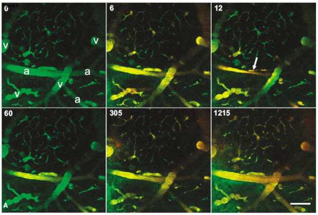
FIGURE 9.
In vivo multiphoton imaging of the mouse brain vasculature during FUS-induced BBB disruption. The animal received 0.1ml (2 mg/ml) 10 kDa, dextran-conjugated Alexa Fluor 488 intravenously ~5 min before imaging (green in images). Immediately after the t=0 frame was taken, a 45-s US exposure was initiated and a 0.1ml bolus (10 mg/ml) of 70 kDa, dextran-conjugated Texas Red was delivered intravenously (red in images). Almost total occlusion of the large vessel in the center of the field occurred 12 s after the initiation of ultrasound exposure (arrow). Beginning at 60 s and by 305 s, leakage in the green channel is apparent in the lower left of the field, and around the central vessel. (a=arterioles; v=veins; scale bar is 100 μm). Reproduced with permission from [204].

