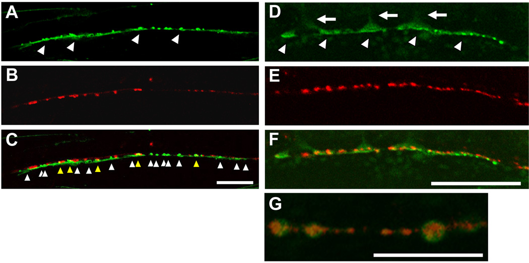
Figure 8. Synaptic localization of Na+/K+ ATPase and the distinct localization of the nAchR.
(A) Localization pattern of the NKB-1::GFP fusion protein around the anterior ventral nerve cord. The fusion protein formed a punctate pattern along the ventral nerve cord. (B) Localization pattern of the presynaptic SNG-1::mRFP fusion protein expressed under the control of the unc-25 promoter. The puncta indicate GABAergic presynaptic sites. (C) Merged image. Although several of the mRFP and GFP puncta were colocalized (yellow arrowhead), a lot of GFP puncta did not colocalize with mRFP (white arrowhead), suggesting a predominant localization of the Na+/K+ ATPase at cholinergic postsynaptic sites. Scale bars, 20 µm. (D) Localization pattern of the NKB-1::GFP fusion protein. Arrowheads indicate the NMJs. Arrows indicate the muscle arms innervated from the muscle cells. (E) Localization pattern of the LEV-1:: 3xHA fusion protein. LEV-1 is a nAchR non-α subunit expressed in body-wall muscles, and membrane-inserted LEV-1 protein was visualized by staining with Alexa-594-labelled anti-HA antibodies. (F) Merge image. Scale bars, 20 µm. (G) In some cases, nAchR clusters (UNC-38:: myc, red) were surrounded by Na+/K+ ATPase (EAT-6:: GFP, green). Scale bars, 10 µm. All pictures show the ventral view.

