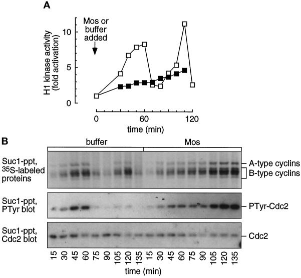
Figure 6.
Cyclins accumulate and associate with Cdc2 in MAP kinase-activated extracts. Cycling extracts were supplemented with sperm chromatin, [35S]methionine, and buffer (□) or Mos (▪). (A) Histone H1 kinase assays were performed on samples removed every 10 min. (B) Cyclin accumulation and Cdc2 tyrosine phosphorylation. Parallel samples were removed every 15 min and subjected to p13suc1 agarose precipitation and SDS-polyacrylamide gel electrophoresis. Suc1-precipitated 35S-labeled proteins were detected by autoradiography (upper). The cyclins exhibit an abrupt decrease in signal at mitosis (75 min) in the buffer-treated extracts while continuing to accumulate in the Mos-treated extracts. The lower half of the blot was probed with a phosphotyrosine antibody (middle), stripped, and reprobed with a Cdc2 antibody (lower). The level of phosphotyrosine increases over time in the Mos-treated extracts, and cycles in the buffer-treated extracts. Levels of cyclin, Cdc2, and Cdc2-PTyr were quantified by PhosphorImager scanning.

